Home
/ Loculated Pleural Effusion Cxr : Pleural Effusion / What does pleural effusion mean?
Loculated Pleural Effusion Cxr : Pleural Effusion / What does pleural effusion mean?
Loculated Pleural Effusion Cxr : Pleural Effusion / What does pleural effusion mean?. The cardiac silhouette is also obscured. Dr bhatia discussing on pleural effusion in #lastminuterevisionpointdiscussionseries. Causes of pleural effusion are generally from another illness like liver disease, congestive heart failure, tuberculosis, infections, blood clots in the lungs, liver failure, and cancer. Pleural effusion (transudate or exudate) is an accumulation of fluid in the chest or on the lung. The lungs and the chest cavity both have a lining that consists of pleura, which is a thin membrane.
Transudates or exudates as defined by lights criteria. Pleural fluid/serum ldh ratio >0.6. Pleural effusion occurs when too much fluid collects in the pleural space (the space between the two layers of the pleura). e intrinsic characteristics of an effusion and its. no change in position of effusion withchange in position of chest.
Radiografía torácica: Derrames pleurales - UpToMedicine from images.radiopaedia.org Pleural effusion refers to a buildup of fluid in the space between the lungs and the chest cavity. Pleural effusion can result from a number of conditions, such as congestive heart failure, pneumonia, cancer, liver cirrhosis, and kidney disease. Determine if it can be tapped. produced at parietal and resorbed atvisceral pleura. Among the causes, pleural infection, heart failure, and malignan. Blunting of costophrenic angle initially. There is always a small amount of fluid around the lung t. Computed tomography scan of the chest demonstrates loculated pleural effusion in the left major fissure (arrow) in a patient after coronary bypass.
Occasionally, a focal intrafissural fluid collection may look like a lung mass.
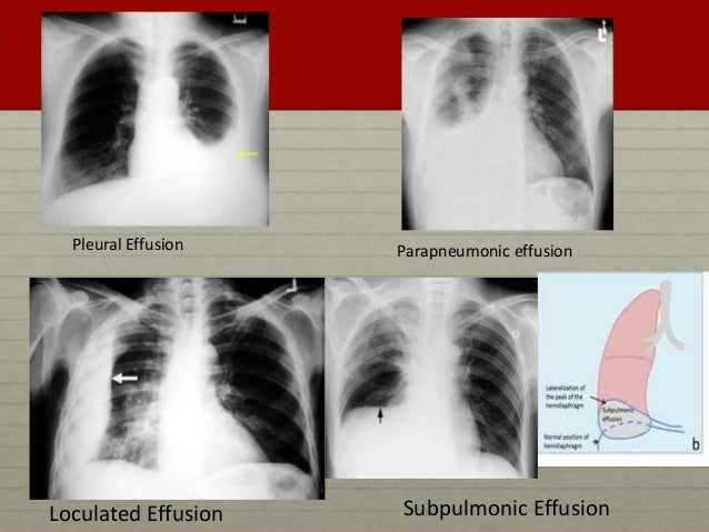
What are the pulmonary findings? Dr bhatia discussing on pleural effusion in #lastminuterevisionpointdiscussionseries. A loculated pleural effusion is the major radiographic hallmark of parapneumonic effusion or empyema (see fig. Pleural fluid ldh > two thirds of upper limit for serum ldh. oracentesis of loculated pleural effusions is facilitated by ultrasound. Pleural effusions can loculate as a result of adhesions. There is a large left pleural effusion obscuring the lower half of the left hemi thorax. Among the causes, pleural infection, heart failure, and malignan. The pleura is a thin membrane that lines the surface of your lungs and the inside of your chest wall. Differentiation of loculated effusions from solid masses. A pleural effusion is accumulation of excessive fluid in the pleural space, the potential space that surrounds each lung. Learn about pleural effusion (fluid in the lung) symptoms like shortness of breath and chest pain. The theory is that a local pleuritis causes the pleura to thicken and contract.
Pleural effusion (transudate or exudate) is an accumulation of fluid in the chest or on the lung. The cardiac silhouette is also obscured. The theory is that a local pleuritis causes the pleura to thicken and contract. Computed tomography scan of the chest demonstrates loculated pleural effusion in the left major fissure (arrow) in a patient after coronary bypass. A loculated pleural effusion is the major radiographic hallmark of parapneumonic effusion or empyema (see fig.
Pleural Effusion for Undergraduates from image.slidesharecdn.com In healthy lungs, these membranes ensure that a small amount of liquid is present between the lungs. The underlying lung shrinks and atelectasis develops in a round configuration. When you have a pleural effusion, fluid builds up in the space between the layers of your pleura. Pleural effusions are a common medical problem with more than 50 recognised causes including disease local to the pleura or underlying lung, systemic conditions, organ dysfunction and drugs.1. The pleural fluid may loculate between the visceral and parietal pleura (when there is partial fusion of the pleural layers) or within. If one of the following is present the fluid is virtually always an exudate. Blunting of costophrenic angle initially. Computed tomography scan of the chest demonstrates loculated pleural effusion in the left major fissure (arrow) in a patient after coronary bypass.
Pleural effusion occurs when too much fluid collects in the pleural space (the space between the two layers of the pleura).
Meniscus sign is a rim of fluid ascending the lateral chest wall. Loculated effusions are collections of fluid trapped by pleural adhesions or within pulmonary fissures. Other causes are complicated parapneumonic effusion. Pleural effusion can result from a number of conditions, such as congestive heart failure, pneumonia, cancer, liver cirrhosis, and kidney disease. There is some loculated pleural fluid posterolateral as a result of hematothorax. Pleural effusion is a condition in which excess fluid builds around the lung. Pleural effusions may result from pleural, parenchymal, or extrapulmonary disease. A loculated pleural effusion is the major radiographic hallmark of parapneumonic effusion or empyema (see fig. Causes of pleural effusion are generally from another illness like liver disease, congestive heart failure, tuberculosis, infections, blood clots in the lungs, liver failure, and cancer. Mediastinal shift (tracheal deviation) if fluid levels >1000mls. Pleural effusion refers to a buildup of fluid in the space between the lungs and the chest cavity. Approximately 1 million people develop this abnormality each year in the united states. Blunting of costophrenic angle initially.
The underlying lung shrinks and atelectasis develops in a round configuration. Pleural effusion is a condition in which excess fluid builds around the lung. Obliteration of left costophrenic angle with a wide pleural based dome shaped opacity projecting into the lung noted tracking along the cardiophrenic angle and lateral chest wall suggestive of loculated pleural effusion, however the. My pleural effusion healed without treatment. Empyema is defined as the presence of pus in the pleural space.
The patient's chest radiograph (CXR)-large-sized, right ... from www.researchgate.net Among the causes, pleural infection, heart failure, and malignan. Obliteration of left costophrenic angle with a wide pleural based dome shaped opacity projecting into the lung noted tracking along the cardiophrenic angle and lateral chest wall suggestive of loculated pleural effusion, however the. Accompanying adhesions can be identified. Blunting of costophrenic angle initially. e intrinsic characteristics of an effusion and its. 9 633 просмотра 9,6 тыс. If none is present the fluid is virtually always a transudate. Occasionally, a focal intrafissural fluid collection may look like a lung mass.
Pleural fluid/serum ldh ratio >0.6.
Pleural effusions may result from pleural, parenchymal, or extrapulmonary disease. Pleural effusions are a common medical problem with more than 50 recognised causes including disease local to the pleura or underlying lung, systemic conditions, organ dysfunction and drugs.1. Occasionally, a focal intrafissural fluid collection may look like a lung mass. It is commonly known as water on the lungs. Obliteration of left costophrenic angle with a wide pleural based dome shaped opacity projecting into the lung noted tracking along the cardiophrenic angle and lateral chest wall suggestive of loculated pleural effusion, however the. Pleural effusions occur as a result of increased fluid formation and/or reduced fluid resorption. When you have a pleural effusion, fluid builds up in the space between the layers of your pleura. e intrinsic characteristics of an effusion and its. 9 633 просмотра 9,6 тыс. My pleural effusion healed without treatment. Accompanying adhesions can be identified. Pleural effusion occurs when too much fluid collects in the pleural space (the space between the two layers of the pleura). Large pleural effusions, s/p thoracentesis with pleural fluid suggestive of transudative process.
Pleural effusion refers to a buildup of fluid in the space between the lungs and the chest cavity loculated pleural effusion. The theory is that a local pleuritis causes the pleura to thicken and contract.